
The Canine Chest in 3D: Lungs, Vessels, Heart and Airways
Here's a walkthrough of our canine chest cavity content. 🫁 It's split into four scenes, covering lung lobation, the vessels of the chest, the circulations of the heart and the bronchial tree.
Scene 1: lung fields
The first scene shows the lungs split into their different lung fields. You can select them and click through them, and hitting the space bar splits them apart. You can also hide them individually in the normal way, with the H key or the menu on the side.
Scene 2: vasculature of the chest
The second scene covers the major vessels within the chest cavity, such as the cranial vena cava and the aorta. You can hide and show as many or as few as you like, and use the search function to find the vessels you want to look at.
Scene 3: the circulations from a CT scan
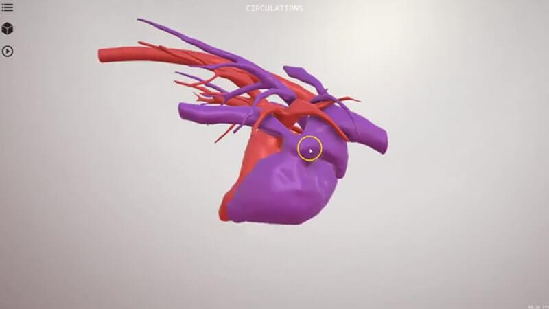
The third scene was made from a CT scan. It shows the left side and the right side of the circulation, giving you an idea of what the atria and ventricles look like within the myocardium and how they contract. We've also marked approximately where the SA and AV nodes reside, which is a nice anchor when you're thinking about conduction.
Scene 4: the bronchial tree
The last scene is the bronchial tree, the airways of the dog, shown in a right lateral view. Again, you can use the space bar to split it apart into the different bronchi, which is a great way to get to grips with the anatomy before you look at thoracic radiographs.
Explore more
FREE for veterinary students, interns, residents & academics at vin.com. Also available by subscription at ivalalearn.com
Want to understand more about the options for accessing the content? Our company FAQs have the answers here pricing, account access and billing.



