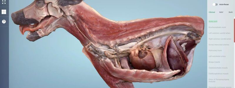
Canine Cranial Hemisection in 3D, Scanned From a Real Specimen
A beautiful scanned dissection of the cranial half of a dog, showing the thorax and cranial abdomen in situ plus the superficial muscles on the other side.

Ivalalearn
Proud to be part of the Veterinary Information Network family

A beautiful scanned dissection of the cranial half of a dog, showing the thorax and cranial abdomen in situ plus the superficial muscles on the other side.
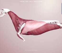
The body wall muscles are a tricky area to picture. Our 3D scene shows how the layers sit in relation to one another, with information on every muscle.
Ivalalearn
Proud to be part of the Veterinary Information Network family
OUR MISSION:
To shape more confident students and clinicians by creating intuitive, engaging, interactive learning content.
OUR MISSION:
To shape more confident students and clinicians by creating intuitive, engaging, interactive learning content.
Ivala is proud to be a wholly owned subsidiary of the Veterinary Information Network (VIN)
IVALA ® All rights reserved.



