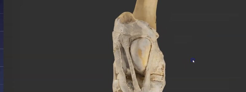
Equine Stifle in 3D, Scanned From a Real Dissection
A scan of a real dissected equine stifle, one of the most interesting areas of musculoskeletal anatomy and home of the locking mechanism.

Ivalalearn
Proud to be part of the Veterinary Information Network family

A scan of a real dissected equine stifle, one of the most interesting areas of musculoskeletal anatomy and home of the locking mechanism.
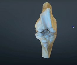
Our scanned, flexed bovine stifle joint adds the distal femur to the proximal tibia, so you can learn the ligaments and how the cruciates attach.
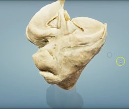
A scan of the proximal tibia of a cow, which our anatomists say is extremely similar to the horse, to help you understand the ligaments of the stifle.
Ivalalearn
Proud to be part of the Veterinary Information Network family
OUR MISSION:
To shape more confident students and clinicians by creating intuitive, engaging, interactive learning content.
OUR MISSION:
To shape more confident students and clinicians by creating intuitive, engaging, interactive learning content.
Ivala is proud to be a wholly owned subsidiary of the Veterinary Information Network (VIN)
IVALA ® All rights reserved.



