
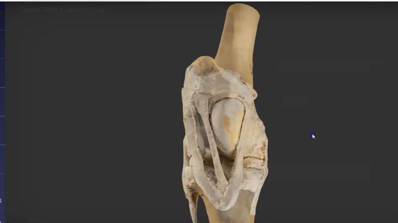
Equine Stifle in 3D, Scanned From a Real Dissection
We've just made available the equine stifle joint, a scan of a real dissected specimen. 🐴 The equine stifle is perhaps one of the most singularly interesting areas of musculoskeletal anatomy, and it's very different in its makeup and structure from the stifle of smaller mammals.
Home of the locking mechanism
The horse's stifle is famous for its locking mechanism, which allows horses to stand for long periods with minimal muscular effort. In this content you can explore some of the structures involved in making that possible. For the bigger picture, see our equine hindlimb stay apparatus.
For anatomy, surgery and arthroscopy
We think it'll be a great learning tool not only for those learning anatomy, but also for anyone performing procedures in the area, such as surgery and arthroscopy. As it's a scan, you can't hide structures, but you can click on them for more information, and the fidelity and accuracy of the model are extremely high. Interested in comparative anatomy? Our bovine proximal tibia is remarkably similar.
Where to find it
Follow the new icon in the menu. On IVALA Learn, it's in the 3D Anatomy Viewer. On VIN, use the Student Center or Library > Learn Anatomy in 3D.
Explore more
FREE for veterinary students, interns, residents & academics at vin.com. Also available by subscription at ivalalearn.com
Want to understand more about the options for accessing the content? Our company FAQs have the answers here pricing, account access and billing.



