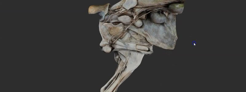
Canine Male Urogenital Tract in 3D, Scanned From a Dissection
A scan of the male canine urogenital tract, with organs and nerves on one side and the caudal body wall and hindlimb muscles on the other.

Ivalalearn
Proud to be part of the Veterinary Information Network family

A scan of the male canine urogenital tract, with organs and nerves on one side and the caudal body wall and hindlimb muscles on the other.
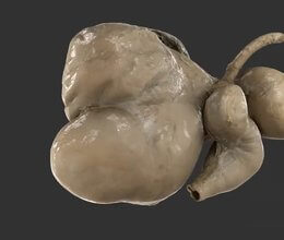
A scan of the ruminant stomachs reconstructed from a real specimen, adding another level of realism to our CT-based schematic.
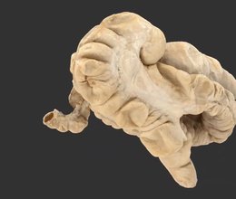
We've added a scan of the equine large intestine from a real specimen, with labelled sacculations and taenial bands, to complement our schematic.
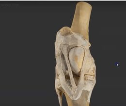
A scan of a real dissected equine stifle, one of the most interesting areas of musculoskeletal anatomy and home of the locking mechanism.
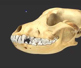
We've rebuilt our canine skull landmarks from a scan of a real skull, so you now select whole anatomical regions instead of small dots.
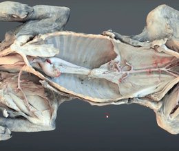
A scanned dissection of a dog revealing the major arterial circulation in situ, from the neck all the way to the median sacral artery.
Ivalalearn
Proud to be part of the Veterinary Information Network family
OUR MISSION:
To shape more confident students and clinicians by creating intuitive, engaging, interactive learning content.
OUR MISSION:
To shape more confident students and clinicians by creating intuitive, engaging, interactive learning content.
Ivala is proud to be a wholly owned subsidiary of the Veterinary Information Network (VIN)
IVALA ® All rights reserved.



