
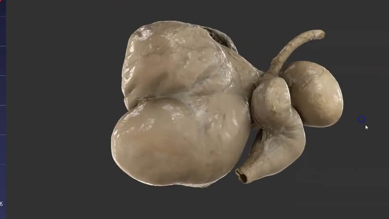
Ruminant Stomachs in 3D, Scanned From a Real Specimen
We're also releasing a scan of the ruminant stomachs. 🐄🐑🐐 It's arguably the most important region of ruminant anatomy to understand.
Another level of realism
We already have schematics of the ruminant stomachs that were made from CT. This scan, though, has been reconstructed from a real anatomical specimen, so the quality and fidelity are that much higher and more lifelike (and more beautiful 💅🏼). It adds another level of realism that can really help with visualisation.
Hover and click for information
Just like our other content, you can hover and click on the different areas to get anatomical information about them. The reticulum card, for example, explains how it mechanically separates particles of digesta, and how its mucosal folds resemble a honeycomb.
Where to find it
On IVALA Learn, it's in the 3D Anatomy Viewer. On VIN, you'll find the Anatomy Viewer on the Learn Anatomy in 3D page.
Explore more
FREE for veterinary students, interns, residents & academics at vin.com. Also available by subscription at ivalalearn.com
Want to understand more about the options for accessing the content? Our company FAQs have the answers here pricing, account access and billing.



