
Equine Forelimb From the Elbow Down: Veins, Arteries and Nerves
A scan of the equine forelimb from roughly the elbow down, dissected to reveal the veins, arteries and nerves, colourised to make them easier to see.

Ivalalearn
Proud to be part of the Veterinary Information Network family

A scan of the equine forelimb from roughly the elbow down, dissected to reveal the veins, arteries and nerves, colourised to make them easier to see.
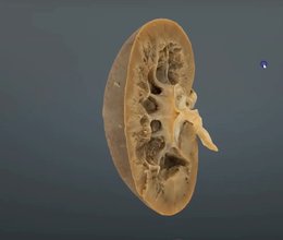
A scanned cut section through the canine kidney, showing the capsule, cortex, medulla and other core structures.
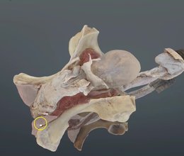
Our scan of the stallion's reproductive tract shows where the accessory sex glands sit and makes sense of the prepuce and its layers.
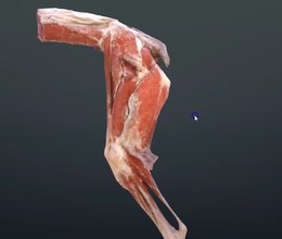
A scan of the canine hindlimb muscles reconstructed from a real cadaver, showing how the muscle bellies are really laid out.
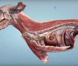
A beautiful scanned dissection of the cranial half of a dog, showing the thorax and cranial abdomen in situ plus the superficial muscles on the other side.
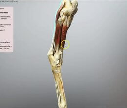
The muscles of the equine distal hindlimb, scanned from a real limb, to give you a much clearer picture of how the flexors and extensors relate.
Ivalalearn
Proud to be part of the Veterinary Information Network family
OUR MISSION:
To shape more confident students and clinicians by creating intuitive, engaging, interactive learning content.
OUR MISSION:
To shape more confident students and clinicians by creating intuitive, engaging, interactive learning content.
Ivala is proud to be a wholly owned subsidiary of the Veterinary Information Network (VIN)
IVALA ® All rights reserved.



