
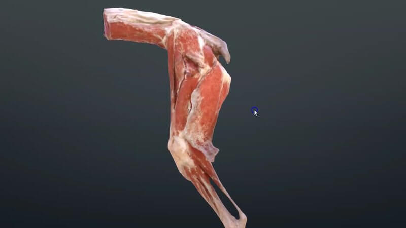
Canine Hindlimb Muscles in 3D, Scanned From a Real Specimen
We've just released a scan of the canine hindlimb muscles. 🐕 It's created from a reconstruction of a real cadaver, so it shows the muscle topography in as close to a real-life way as possible.
How the muscles really look
What's nice about these scans is that they give you a really accurate picture of how the muscles look in real life, compared with the schematic content we already have for the canine hindlimb. The layout of the muscle bellies is much more true to life, which is exactly what you want when you're palpating a limb or planning a surgical approach.
Use it alongside the schematic content
The one drawback of scanned content is that you can't hide the surface layers to look at the deeper ones. That's where our schematic canine hindlimb muscles come in. Used together, we think they're a really powerful way to understand the musculature of the region. We did the same for the front end too, see our canine forelimb muscle scan.
Where to find it
On IVALA Learn, it's in our all-in-one 3D Anatomy Viewer. On VIN, you'll find it on the Learn Anatomy in 3D page, which you can reach from the anatomy interactive content link in the Student Center or via the Library.
Explore more
FREE for veterinary students, interns, residents & academics at vin.com. Also available by subscription at ivalalearn.com
Want to understand more about the options for accessing the content? Our company FAQs have the answers here pricing, account access and billing.



