
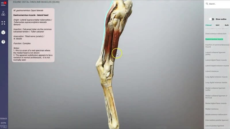
Equine Distal Hindlimb Muscles in 3D, Scanned From a Real Limb
Hot on the heels of the equine forelimb muscles, here are the muscles of the distal equine hindlimb. 🦵🏼
Flexors and extensors, finally clear
The relation of the flexors and extensors on the different parts of the distal limb has always been tricky to understand. We're really hoping this content gives you a much clearer picture of exactly how these muscles are related spatially.
Scanned from a real limb
The content was taken from a real scan of the muscles of a distal equine hindlimb, so the fidelity is super accurate, and you can see just how much detail there is as you spin it around. You can click on the muscles to interact with them, and test yourself using MCQs or by typing the name, directly within the content.
Where to find it
It's part of our equine musculoskeletal project. Open the menu: the first scene you're taken to is the equine skeleton, and the new scene is Equine distal hindlimb muscles. Please use it, enjoy it, learn from it, and let us know what you think.
Explore more
FREE for veterinary students, interns, residents & academics at vin.com. Also available by subscription at ivalalearn.com
Want to understand more about the options for accessing the content? Our company FAQs have the answers here pricing, account access and billing.



