
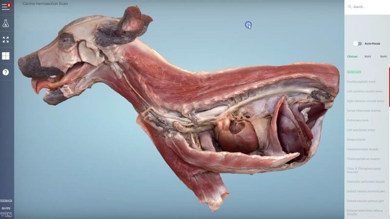
Canine Cranial Hemisection in 3D, Scanned From a Real Specimen
We have some beautiful new content for you: a hemisection through the cranial half of a dog. 🐶 It gives you a really nice look inside the chest and the cranial abdomen, with everything sitting where it belongs.
Real structures in situ
As you hover over the structures they light up, and you can click on any of them for more information. Because this content is made from scans of a real specimen, you get to see fine structures that are really hard to recreate accurately by hand, such as the ansa subclavia. It's an extremely accurate look at the anatomy of this region, about as close as you can get to learning from a real dissection.
Superficial muscles on the other side
Turn the specimen around and you'll find a dissection of the muscles of the region. So it's not only useful for the structures inside the thorax and abdomen, but also for the major superficial musculature of the forelimb and the body wall. Seeing both sides of the same dog helps you connect what you palpate on the outside with what sits underneath, which comes in handy for thoracic auscultation, imaging and surgical approaches.
If you'd like to compare it with our schematic content, have a look at the canine right hemithorax. We've got more great stuff on the way!
Explore more
FREE for veterinary students, interns, residents & academics at vin.com. Also available by subscription at ivalalearn.com
Want to understand more about the options for accessing the content? Our company FAQs have the answers here pricing, account access and billing.



