
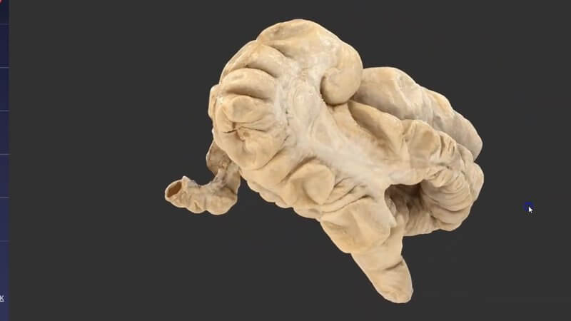
Equine Large Intestine in 3D, Scanned From Real Anatomy
We've added a scan of the large intestinal tract of the horse. 💫🐎 It's a fundamental anatomical region that we've covered before in schematic form, and now we're taking the realism to the next level.
From schematic to real specimen
Our equine abdomen schematic does a great job of showing the general layout of the large intestine alongside the other abdominal organs. Because this scan is created from a real specimen, its quality and accuracy are much higher than anything we can build by hand.
Haustra, sacculations and taenial bands
You can see features such as the haustra and sacculations and the different taenial bands really nicely, and they're all labelled. Hover and click on any area for more information; the card for the taenial bands, for example, reminds you that the caecum has four of them. These are exactly the features you use to orientate yourself on rectal examination or at colic surgery, so it pays to know them well.
Where to find it
It's under Equine abdomen > Large intestine scan, or just follow the new tags in the menu. On IVALA Learn, it's in the 3D Anatomy Viewer. On VIN, go to Library > Learn Anatomy in 3D.
Explore more
FREE for veterinary students, interns, residents & academics at vin.com. Also available by subscription at ivalalearn.com
Want to understand more about the options for accessing the content? Our company FAQs have the answers here pricing, account access and billing.



