
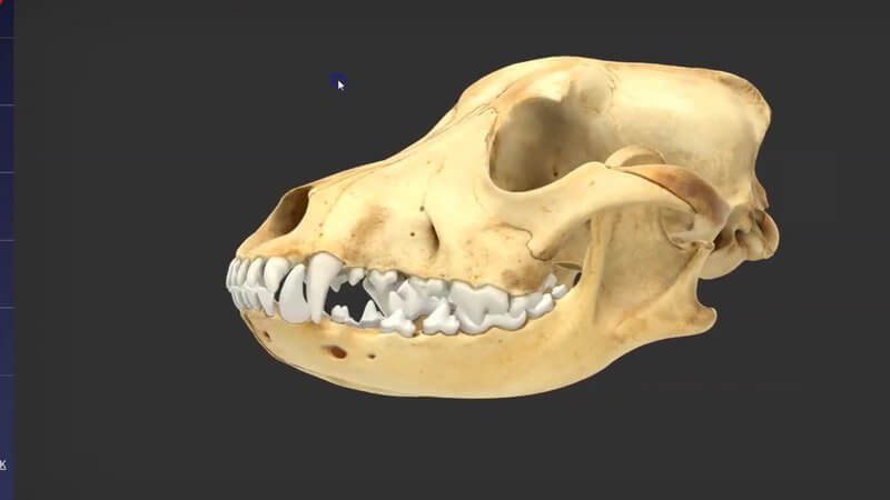
Canine Skull Landmarks in 3D, Refreshed With a Real Scan
We've updated one part of our canine skull content: the bony landmarks of the canine skull. 🦴 This time we used a scan of a real skull, so it's much higher fidelity, more beautiful and a better learning experience all round. 🤗
From dots to real regions
We had skull landmarks before, but it was very old content. It worked, but interacting with the skull through a set of dots was a bit imprecise. The new version highlights the actual regions that are relevant to the anatomy, such as the masseteric fossa (fossa masseterica) and the angular process (processus angularis), with both the English and Latin names.
The old content is staying for now
The new model will eventually replace the old one, but we'll keep the original there for now for anyone who's still using it. If you'd like to compare, here's our post on the original canine skull landmarks, teeth and canals. You'll find the refresh in the usual places: the 3D Anatomy Viewer on IVALA Learn and the 3D content on VIN.
Explore more
FREE for veterinary students, interns, residents & academics at vin.com. Also available by subscription at ivalalearn.com
Want to understand more about the options for accessing the content? Our company FAQs have the answers here pricing, account access and billing.



