
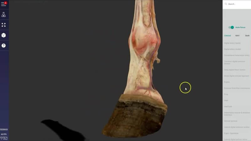
Equine Distal Forelimb Dissection in 3D: Nerves and Vessels
We've added the dissected distal left forelimb of the horse to our equine distal limb (hoof) content. 🐴 It was made from a scan of a real dissected limb, so the quality is really, really high.
Nerves, arteries and veins
This content is extremely useful for studying the nerve supply and arterial supply of the distal limb, as well as its venous drainage.
Made for nerve blocks
It's especially handy if you're studying nerve blocks: the landmarks you need to know for placing your blocks, and why you place them where you do. Seeing the nerves on a real dissection makes those landmarks much easier to remember when you're standing next to a lame horse.
One thing to note
Because it comes from a scan, you can't hide any of the structures. You can only interact with the surface anatomy, but we think that's still going to be extremely useful.
Tell us what you think
Please interact with it, play with it, and if there's anything you'd like us to add, get in touch. We always love receiving your feedback.
Explore more
FREE for veterinary students, interns, residents & academics at vin.com. Also available by subscription at ivalalearn.com
Want to understand more about the options for accessing the content? Our company FAQs have the answers here pricing, account access and billing.



