
Equine Distal Limb: Tendons, Ligaments & Hoof
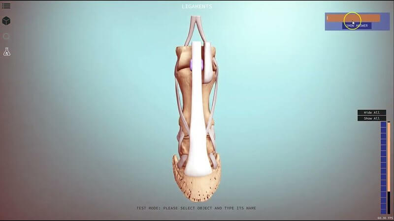
This short clip is a hands-on look at our equine tendons, ligaments and hoof 3D scene - mostly demo, not narration - so you can see how the distal limb hangs together before you open it yourself. 🐴
Equine distal limb tendons and ligaments in 3D
The model centres on the distal limb: cannon region, sesamoids, pastern bones and the hoof capsule, with soft-tissue structures you can select. On-screen info in the video highlights the interosseous muscle and extensor branches (aka suspensory ligament) - described as a predominantly tendinous band with origin from the palmar carpal ligament and metacarpal bone - plus labels such as DDFT, common digital extensor, and various palmar ligaments.
Hide, show and Test Mode
Use the structure list to hide and show objects, isolate bones versus soft tissue, and enter Test Mode: select an object, type its name, and use Show Answer when you need a nudge. 🎯
Explore more
FREE for veterinary students, interns, residents & academics at vin.com. Also available by subscription at ivalalearn.com
Want to understand more about the options for accessing the content? Our company FAQs have the answers here pricing, account access and billing.



