
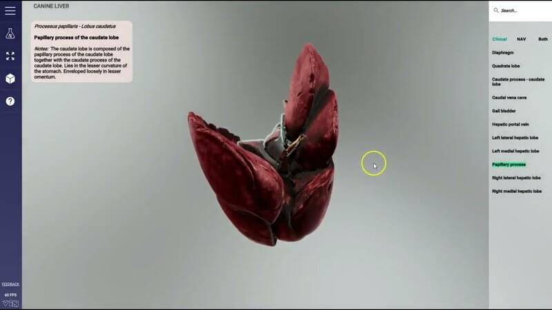
Canine Liver in 3D from a Real Organ Scan
Our new canine liver 3D model comes from scanning a real organ - not just CT or MRI. 🫀 Previously we rebuilt organs from imaging; here we take real organs postmortem, scan them with different techniques, and reconstruct their structure as they'd be found in situ.
Canine liver detail you can almost feel
The level of structural detail is next-level. For instance, you can pick out the caudate process of the caudate lobe and see the recess for the kidney - so you really appreciate how this organ sits in situ.
What changed in the UI for scanned organs
Because of the scanning process and the organ, we can't hide different structures individually the way we do in other projects. You can still select structures to see what they are, but you can't hide them, and the checkboxes on the right are gone. Everything else is the same: click to select and get info, click elsewhere to deselect.
Test Yourself still works
Test Yourself modes work as before - worth trying if you haven't. In the walkthrough we check a label such as left medial hepatic (and yes, we had to look that one up too). 😄
New in-content feedback form
We've added a feedback form inside the content: click feedback, add your name, email and message, and we'll get the email - and we promise we'll respond. Especially if it's bad feedback. We want that.
Explore more
FREE for veterinary students, interns, residents & academics at vin.com. Also available by subscription at ivalalearn.com
Want to understand more about the options for accessing the content? Our company FAQs have the answers here pricing, account access and billing.



