
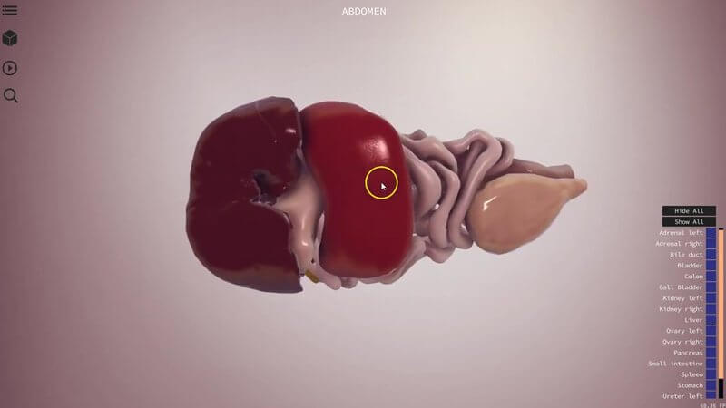
The Canine Abdomen in 3D, Plus a Radiography Scene
Here's a quick walkthrough of our canine abdomen scene. 🐕 We've extracted all of the organs from a dog CT, so it's very accurate when it comes to understanding organ relations and the topography of the abdominal cavity.
Organs exactly where they lie
You can hide the rib cage or keep it in view as a landmark. The most useful part is starting to understand the spatial relations between organs, for instance which structures you'll come across when you're entering the abdomen during surgery and roughly how they lie. This one is a female dog, so you'll see the uterus, and we've included the adrenal glands too.
A radiography scene
The project also includes a radiography scene. Here we've made all the organs always visible as outlines, so you can always get an idea of their shape and position, a bit like on an X-ray.
That's really handy when you're taking a right lateral, left lateral or a ventrodorsal view. You can see, for example, how the colon lies, and get a better feel for where each structure should be and where you'll see it on the radiograph.
Have a play
Have a look, play around and let us know your thoughts.
Explore more
FREE for veterinary students, interns, residents & academics at vin.com. Also available by subscription at ivalalearn.com
Want to understand more about the options for accessing the content? Our company FAQs have the answers here pricing, account access and billing.



