
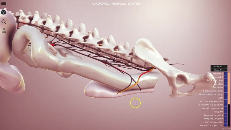
The Canine Autonomic Nervous System in 3D
The autonomic nervous system is one of those topics that's notoriously hard to picture. 🧠 This is a preview of our canine autonomic nervous system 3D content, where we set out to provide a schematic of the system in the dog, so you can understand the spatial relationships and course of its sympathetic and parasympathetic parts.
Sympathetic and parasympathetic, colour coded
The sympathetic system is shown in black, so you can start to appreciate its thoracolumbar outflow from the spinal cord. The parasympathetic system is in orange, with its craniosacral outflow.
Interactive markers along the way
The red markers you can see along the system are all interactive. Click one and it tells you about that structure. For instance, there's a marker on the pelvic nerve, which contains fibres from the parasympathetic system. You'll also find the different ganglia, such as the left coeliac ganglion, along with the organs they're closely related to.
Why a schematic helps
Gaining a good understanding of this system is very difficult, because you can't really dissect it out intact. That makes it hard to appreciate the relations of all the nerves as a whole. By reviewing the outflows in 3D, we hope you'll come away with a much clearer picture, which pays off whenever autonomic function comes up in physiology, pharmacology or the clinic.
Let us know your thoughts
Check it out and tell us what you think. We'd love to hear how you use it.
Explore more
FREE for veterinary students, interns, residents & academics at vin.com. Also available by subscription at ivalalearn.com
Want to understand more about the options for accessing the content? Our company FAQs have the answers here pricing, account access and billing.



