
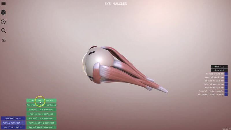
Canine Eye Muscles in 3D: Animated Function and Nerve Lesions
We've added a new scene to our canine eye content: the extrinsic musculature of the canine eyeball. 👀 As a reminder, you can switch between the different scenes in a project using the menu in the top left corner.
Why the eye muscles?
It's quite a niche topic, but it served as a really good test bed for us to see how animation performs in our 3D content.
Muscle function, animated
In the bottom left corner there's a menu called Muscle function. Choose a muscle and you can see what it does when it contracts, animated right in the scene.
Simulated nerve lesions
Anatomy and function are really important, but that importance stems from the fact that they have clinical relevance. So there's also a Nerve lesions menu, where we can simulate what happens with a lesion of a nerve supplying the eye.
For example, we can simulate a lesion of the oculomotor nerve. It supplies a number of the muscles you can see in the scene, as well as the muscle of the upper eyelid, and with a lesion you get a ventrolateral strabismus and a dilated pupil.
More context to come
We want to give you as much context and relevance in our content as we can, so expect to see more of this kind of animation and pathology going forward. A big thank you again to Ben Blacklock, veterinary ophthalmologist at Dick White Referrals, for his input on this project.
Explore more
FREE for veterinary students, interns, residents & academics at vin.com. Also available by subscription at ivalalearn.com
Want to understand more about the options for accessing the content? Our company FAQs have the answers here pricing, account access and billing.



