
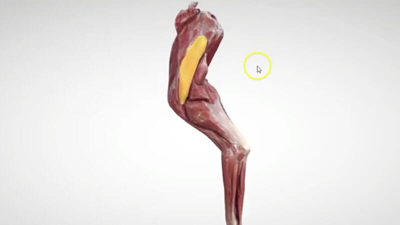
Canine Forelimb Muscles in 3D, Scanned From a Real Specimen
New 3D content: we've added a scan of the canine forelimb muscles to the Superproject. 🐕 We already have a schematic of the forelimb muscles, but this one was created from a scan of a real specimen.
Why a scan makes a difference
Because it comes from a real limb, the accuracy of the muscle origins and insertions is really, really good. It's a much more accurate representation of the muscles, which you'll especially appreciate in the antebrachial area. It's excellent for understanding the flexor and extensor muscle groups, where they originate and bunch up, and how things look in the real limb.
Where to find it
Open the Superproject and go to Canine forelimb, where you'll find the new scanned specimen.
Select, but not hide
Just a reminder: because this content is a scan, you can't hide the muscles, you can only select them. If you want to hide muscles and see the deeper layers, the schematic canine forelimb musculature is still there in the same section, so you can use the two side by side.
Explore more
FREE for veterinary students, interns, residents & academics at vin.com. Also available by subscription at ivalalearn.com
Want to understand more about the options for accessing the content? Our company FAQs have the answers here pricing, account access and billing.



