
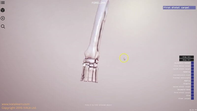
Canine Limb Bones and Landmarks in 3D
Knowing your bony landmarks is the foundation for so much: palpation, radiograph reading, surgical approaches and understanding where muscles attach. 🦴 This scene covers the bones of the canine forelimb and hindlimb, along with their prominences and landmarks.
Select bones and explore the landmarks
You interact with the bones in the normal way: select a bone to see what it is. We've also added markers on particular aspects of the bones that point out landmarks and give you a bit more information. For example, the marker on the coracoid process notes that it's the origin of the coracobrachialis muscle.
The carpus, split apart
Something you might find of particular interest is the carpus, as it's a pretty complex area. All the carpal bones are there, and you can split them apart to see each one on its own. In the video we use the space bar, but the play button does the same thing.
Pelvis and tarsus too
The hindlimb is included as well. You can split the pelvis apart, then come down and look at the tarsus in more detail.
Explore more
FREE for veterinary students, interns, residents & academics at vin.com. Also available by subscription at ivalalearn.com
Want to understand more about the options for accessing the content? Our company FAQs have the answers here pricing, account access and billing.



