
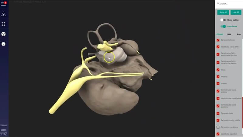
The Canine Middle and Inner Ear in 3D, From a MicroCT Scan
We're releasing something really, really cool: the canine middle and inner ear in 3D. 👂🏼 As far as we're aware, we've never seen a 3D reconstruction of this region in this much detail.
Built from a microCT scan
The reconstruction was made from a microCT scan, which is what gives it such fine detail. It starts with a cut section of the canine skull. Hide that, and you see the internal structures: the nerves in yellow, and the bony structures and spaces that form the cavities of the middle and inner ear.
Structures to look out for
You'll start to recognise things like the external acoustic meatus and the tympanic membrane. Keep hiding and going deeper and you can even see one of the auditory ossicles, as well as a muscle of the region.
Follow the nerves
Bring back the cut section of skull and you can see the internal acoustic meatus, with the vestibular and cochlear nerves entering it, and the facial nerve travelling into the facial canal. Being able to see the passage of all the different nerves through this region is one of the best things about this content.
Why it matters clinically
Seeing how close these nerves run helps explain why middle ear disease is very different from external ear disease, and why it can cause the neurological changes you quite commonly see when animals have middle ear disease.
Let us know what you think
We're really excited to have this content available for you. Please use it, enjoy it and let us know what you think.
Explore more
FREE for veterinary students, interns, residents & academics at vin.com. Also available by subscription at ivalalearn.com
Want to understand more about the options for accessing the content? Our company FAQs have the answers here pricing, account access and billing.



