
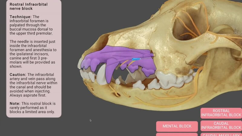
Canine Dental Nerve Blocks in 3D, With Animated Blocks
Dental procedures in dogs are commonplace, yet effective pain management through regional anaesthesia can sometimes be overlooked. Our new canine dental nerve blocks content gives you a clear understanding of injection sites and the areas each block covers, so your patients get optimal pain relief. 🐶🦷
Click a block, watch the animation
It's a little different from our usual anatomy content. In the bottom right corner there are buttons for each block, including the rostral infraorbital, caudal infraorbital, caudal maxillary, mental and mandibular blocks. Click one and a short animation shows the area that's anaesthetised when you place a local block in that location.
Technique notes and cautions
Each block comes with further information on the technique. For the rostral infraorbital block, for example, you palpate the infraorbital foramen through the buccal mucosa, and there's a reminder that the infraorbital artery and vein run with the nerve, so always aspirate first. It makes a handy reference to check before you perform regional anaesthesia.
The nerves that matter
You can also interact with the nerves relevant to dental blocks, such as the maxillary nerve, a branch of the trigeminal nerve, and read about their course and what they supply. You'll find it under Canine > Head & neck > Skull > Dental nerve blocks, and on VIN in the 3D Anatomy Viewer via Library > Learn Anatomy in 3D.
Explore more
FREE for veterinary students, interns, residents & academics at vin.com. Also available by subscription at ivalalearn.com
Want to understand more about the options for accessing the content? Our company FAQs have the answers here pricing, account access and billing.



