
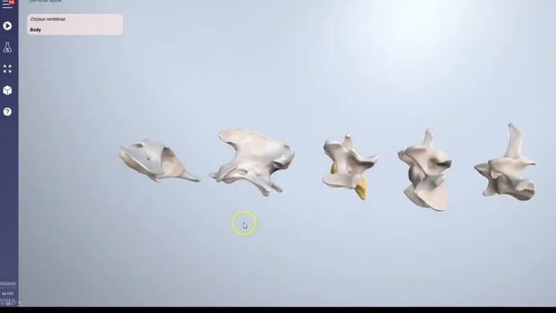
Canine Spine in 3D: Vertebrae and Landmarks, Refreshed
We've refreshed our canine spine content. 🦴 It covers the bones of the cervical, thoracic, lumbar and caudal spine, along with their prominences and landmarks, and now it looks better than ever.
Curious how it looked before the refresh? Here's our original preview:
Higher quality scan data
The first change is that we're now using higher quality scan data, so the surface features of all the different bones are much closer to real life than in the content we used before.
Whole regions instead of buttons
The second big change is how we show the landmarks. Before, we used small buttons, for example to represent the wings of the atlas. Now you can highlight the entire region of each relevant area of anatomy, which makes the landmarks much more obvious. You can look through all the different vertebrae that have defining features.
A typical thoracic and lumbar vertebra
We've also individualised a thoracic vertebra and a lumbar vertebra, so you can look at the features of each one on its own. These are the typical features you'll find across all thoracic vertebrae and across all lumbar vertebrae, so they're a great place to start before you compare regions.
Where to find it
You'll find the spine content under Canine axial region. It's well worth knowing your vertebral landmarks, whether you're reading spinal radiographs or thinking about where to place a needle for an epidural.
Explore more
FREE for veterinary students, interns, residents & academics at vin.com. Also available by subscription at ivalalearn.com
Want to understand more about the options for accessing the content? Our company FAQs have the answers here pricing, account access and billing.



