
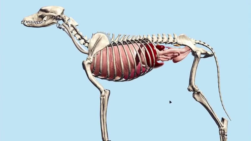
Canine Internal Organs in 3D: Thorax and Abdomen Together
We've released a new scene called Canine Internal Organs. 🐶 It brings together anatomy we already had, the abdominal organs and the thoracic organs, and places them into a single animal.
New connecting structures
We've also added the diaphragm, a fundamental connecting structure between the thorax and the abdomen, as well as the trachea and the oesophagus, so you can follow their passage through the body. Hide a few structures and you can look a little deeper.
The big picture
In practice we usually focus on specific regions: you operate on the abdomen, or you take radiographs of the thorax. But it's useful to have a topographical overview of how it all fits together, and that's what this content is for. For a regional deep dive, see our canine abdomen and canine chest posts.
You'll find it in the menu under Canine internal organs, and on VIN via Library > Learn Anatomy in 3D.
Explore more
FREE for veterinary students, interns, residents & academics at vin.com. Also available by subscription at ivalalearn.com
Want to understand more about the options for accessing the content? Our company FAQs have the answers here pricing, account access and billing.



