
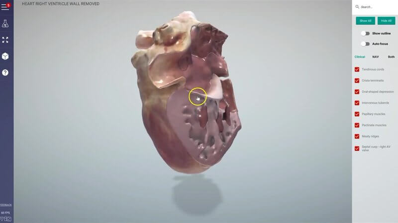
Inside the Canine Heart: Right Ventricle Wall Removed
A very quick update to our canine chest content: the heart with the right ventricular wall removed. 🫀 It lets you look straight inside and explore the structures interactively.
What you can see
With the wall out of the way, you can explore structures such as:
- the papillary muscles
- the chordae tendineae
- the intervenous tubercle
- the fossa ovalis
These are structures that are tricky to picture from a textbook, and a good grasp of them makes it much easier to make sense of what you see on an echo.
Where to find it
You'll find it in our dog chest project, in the third scene, titled Right ventricle wall removed.
Let us know
We hope you enjoy it, and let us know what you think.
Explore more
FREE for veterinary students, interns, residents & academics at vin.com. Also available by subscription at ivalalearn.com
Want to understand more about the options for accessing the content? Our company FAQs have the answers here pricing, account access and billing.



