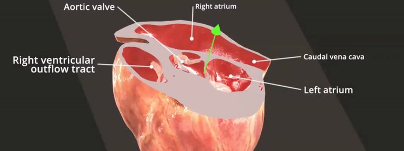
Live Labelling: Labels That Update as You Slice the Heart
Our new live labelling system names anatomical structures on the fly as you slice through the heart, in real time.

Ivalalearn
Proud to be part of the Veterinary Information Network family

These posts cover the thorax, all in the dog. We use 3D models to show the heart, lungs, airways and vessels, and you can slice the heart in any plane or work through a thoracic anatomy MCQ and flashcard set. There is also an echocardiography simulator, so you can practise relating a probe position to the structures in the chest. Other guides look at the right hemithorax, the internal organs of the thorax and cranial abdomen, and live labelling as you section the heart. The material is written for anyone revising thoracic anatomy or preparing for imaging, and it keeps the heart in its wider thoracic context rather than treating it in isolation.
Our new live labelling system names anatomical structures on the fly as you slice through the heart, in real time.
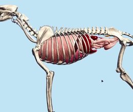
Our Canine Internal Organs scene places the thoracic and abdominal organs in a single animal, with the diaphragm, trachea and oesophagus added.
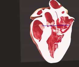
Drag a plane through our 3D canine heart to section it at any position and angle, and explore the chambers and valves inside.
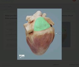
A new set of MCQs on the canine thorax, split into essential and good to know, with instant feedback. You can also use it as a flashcard set.
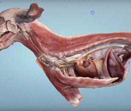
A beautiful scanned dissection of the cranial half of a dog, showing the thorax and cranial abdomen in situ plus the superficial muscles on the other side.
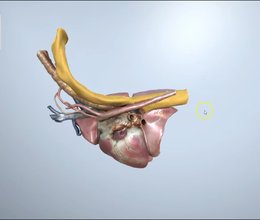
We've added the canine right hemithorax to the Superproject, great for following the oesophagus, and made selected objects highlight in bright orange.
Ivalalearn
Proud to be part of the Veterinary Information Network family
OUR MISSION:
To shape more confident students and clinicians by creating intuitive, engaging, interactive learning content.
OUR MISSION:
To shape more confident students and clinicians by creating intuitive, engaging, interactive learning content.
Ivala is proud to be a wholly owned subsidiary of the Veterinary Information Network (VIN)
IVALA ® All rights reserved.



