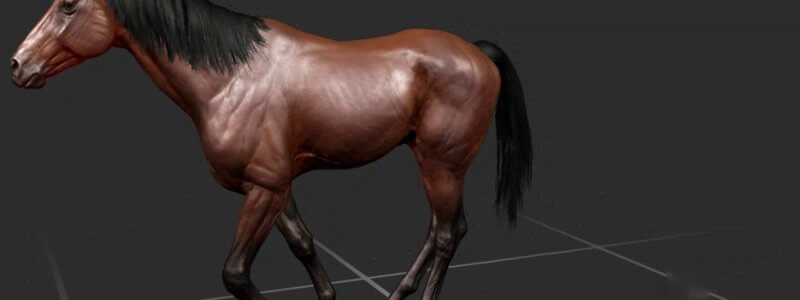
Equine Lameness Gaits in 3D: Grades 1 to 5 at the Walk, Trot and on the Circle
Our animated horse now goes lame: forelimb and hindlimb lameness at grades 1 to 5, at the walk, the trot and on the circle, viewable from any angle.

Ivalalearn
Proud to be part of the Veterinary Information Network family

These posts are about the horse. We use 3D models and animations to show the skeleton, skull and sinuses, distal limb, muscles, nerves, the abdomen and gut, and the stay apparatus. There are also animated gaits, including lameness grades 1 to 5 at the walk, trot and on the circle, so you can practise the observations you would make in a lameness examination. A surface anatomy guide is written to help you use the same language as your clients, and the reproductive anatomy post covers the stallion. The collection is anatomical rather than a complete equine medicine library, but it is organised around the structures and skills that come up often in equine work.
Our animated horse now goes lame: forelimb and hindlimb lameness at grades 1 to 5, at the walk, the trot and on the circle, viewable from any angle.
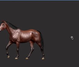
Watch our 3D thoroughbred walk, pace, trot, canter on either lead and gallop from any angle, and slow it right down to study each footfall.
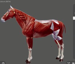
Following our canine full body musculature, the whole musculature of the horse is here, including the trunk and spine muscles we didn't have before.
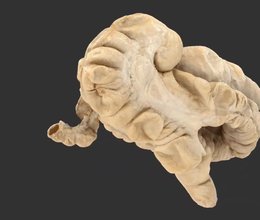
We've added a scan of the equine large intestine from a real specimen, with labelled sacculations and taenial bands, to complement our schematic.
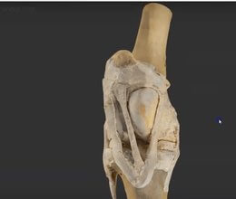
A scan of a real dissected equine stifle, one of the most interesting areas of musculoskeletal anatomy and home of the locking mechanism.
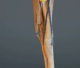
A scan of the equine forelimb from roughly the elbow down, dissected to reveal the veins, arteries and nerves, colourised to make them easier to see.
Ivalalearn
Proud to be part of the Veterinary Information Network family
OUR MISSION:
To shape more confident students and clinicians by creating intuitive, engaging, interactive learning content.
OUR MISSION:
To shape more confident students and clinicians by creating intuitive, engaging, interactive learning content.
Ivala is proud to be a wholly owned subsidiary of the Veterinary Information Network (VIN)
IVALA ® All rights reserved.



