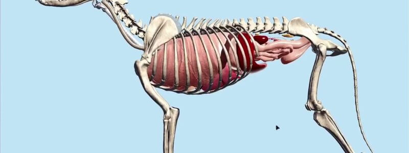
Canine Internal Organs in 3D: Thorax and Abdomen Together
Our Canine Internal Organs scene places the thoracic and abdominal organs in a single animal, with the diaphragm, trachea and oesophagus added.

Ivalalearn
Proud to be part of the Veterinary Information Network family

These posts look inside the abdomen in 3D. We cover organs, vessels and topography in the dog, including the liver, kidney, body wall and a radiography scene, the equine gut, large intestine and male reproductive tract, and the ruminant stomachs. Several guides are written with imaging and surgery in mind, including abdominal vasculature for ultrasound and the body wall muscles relevant to incisions and hernias. You can use the 3D models to sort out organ position and vessel branching before you look at a scan or plan an approach. The collection brings gastrointestinal and urogenital structures together where they sit in the abdomen, rather than splitting them across separate pages.
Our Canine Internal Organs scene places the thoracic and abdominal organs in a single animal, with the diaphragm, trachea and oesophagus added.
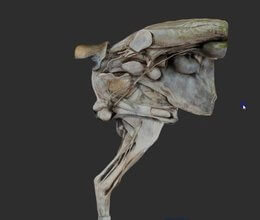
A scan of the male canine urogenital tract, with organs and nerves on one side and the caudal body wall and hindlimb muscles on the other.
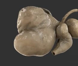
A scan of the ruminant stomachs reconstructed from a real specimen, adding another level of realism to our CT-based schematic.
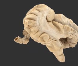
We've added a scan of the equine large intestine from a real specimen, with labelled sacculations and taenial bands, to complement our schematic.
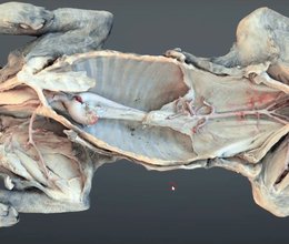
A scanned dissection of a dog revealing the major arterial circulation in situ, from the neck all the way to the median sacral artery.
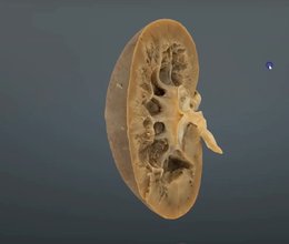
A scanned cut section through the canine kidney, showing the capsule, cortex, medulla and other core structures.
Ivalalearn
Proud to be part of the Veterinary Information Network family
OUR MISSION:
To shape more confident students and clinicians by creating intuitive, engaging, interactive learning content.
OUR MISSION:
To shape more confident students and clinicians by creating intuitive, engaging, interactive learning content.
Ivala is proud to be a wholly owned subsidiary of the Veterinary Information Network (VIN)
IVALA ® All rights reserved.



