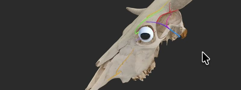
Cow Skull Key Nerves in 3D: Horn and Eye Innervation
Our bovine skull key nerves content highlights the innervation of the horn and eye, a handy review before disbudding, dehorning or eye surgery.

Ivalalearn
Proud to be part of the Veterinary Information Network family

These posts collect 3D anatomy of the head and neck. We cover the skull, teeth, sinuses, brain, eye, ear, larynx and cranial nerves, mainly in the dog and the horse, with a cow skull guide and a few posts on the cat and small mammals. This is useful for dentistry, neurology, ophthalmology and nerve blocks, as well as for everyday revision of landmarks and foramina. Some guides use scanned specimens and microCT, including the canine middle and inner ear and the equine head in median section, so you can see spaces such as the sinuses and guttural pouch more clearly. The posts are grouped here by region, so you can move between adjacent structures rather than jumping between disciplines.
Our bovine skull key nerves content highlights the innervation of the horn and eye, a handy review before disbudding, dehorning or eye surgery.
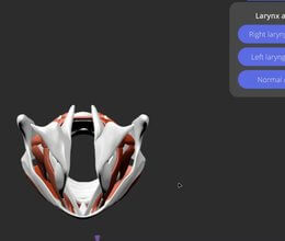
Our canine larynx content demystifies the cartilages and muscles, and animates the larynx during normal respiration and laryngeal paralysis.
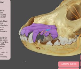
Our canine dental nerve blocks content animates the area each block anaesthetises and adds technique notes, cautions and the relevant nerves.
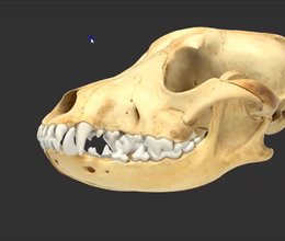
We've rebuilt our canine skull landmarks from a scan of a real skull, so you now select whole anatomical regions instead of small dots.
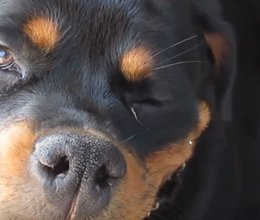
Our vestibular and cerebellar disease deck uses real case videos to help you tell peripheral from central disease and approach each presentation.
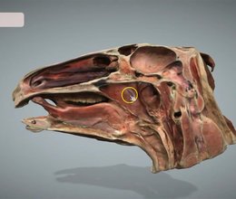
We've added a scanned median section of the equine head, updated the sinus terminology to match the NAV, and made navigation smoother.
Ivalalearn
Proud to be part of the Veterinary Information Network family
OUR MISSION:
To shape more confident students and clinicians by creating intuitive, engaging, interactive learning content.
OUR MISSION:
To shape more confident students and clinicians by creating intuitive, engaging, interactive learning content.
Ivala is proud to be a wholly owned subsidiary of the Veterinary Information Network (VIN)
IVALA ® All rights reserved.



