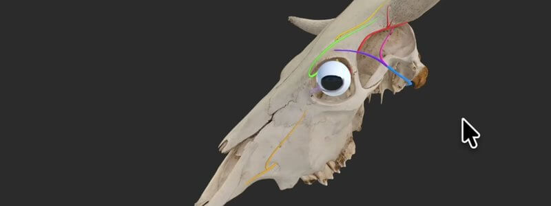
Cow Skull Key Nerves in 3D: Horn and Eye Innervation
Our bovine skull key nerves content highlights the innervation of the horn and eye, a handy review before disbudding, dehorning or eye surgery.

Ivalalearn
Proud to be part of the Veterinary Information Network family

These posts show the nerves you need for regional anaesthesia, in 3D. We cover canine dental blocks and the brachial plexus as it is used for the RUMM block, the veins, arteries and nerves of the equine distal forelimb, and the cornual and ocular innervation of the cow skull, which is relevant to disbudding, dehorning and eye procedures. Each guide is written so you can follow the nerve to the site of the block and see the nearby structures you want to avoid. The material is anatomical rather than a full anaesthetic protocol, but it is organised around the blocks you actually use in practice.
Our bovine skull key nerves content highlights the innervation of the horn and eye, a handy review before disbudding, dehorning or eye surgery.
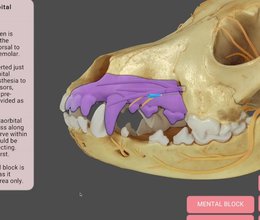
Our canine dental nerve blocks content animates the area each block anaesthetises and adds technique notes, cautions and the relevant nerves.
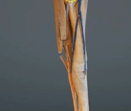
A scan of the equine forelimb from roughly the elbow down, dissected to reveal the veins, arteries and nerves, colourised to make them easier to see.
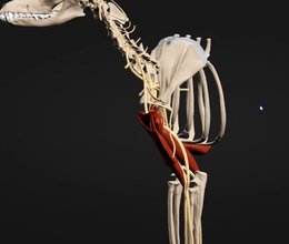
Our canine brachial plexus content was built to help teach the RUMM block, and it's also a great overview of the nerves of the forelimb.
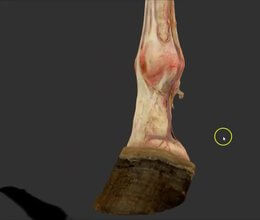
A scan of a real dissected equine distal forelimb, with remarkable detail of the nerves, arterial supply and venous drainage of the limb.
Ivalalearn
Proud to be part of the Veterinary Information Network family
OUR MISSION:
To shape more confident students and clinicians by creating intuitive, engaging, interactive learning content.
OUR MISSION:
To shape more confident students and clinicians by creating intuitive, engaging, interactive learning content.
Ivala is proud to be a wholly owned subsidiary of the Veterinary Information Network (VIN)
IVALA ® All rights reserved.



