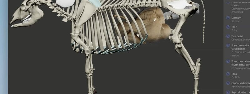
Cow Skeleton in 3D, With the Stomach and Reproductive Tract
We're giving ruminants some extra love with an interactive 3D cow skeleton, with the stomach and reproductive tract added for context.

Ivalalearn
Proud to be part of the Veterinary Information Network family

These posts cover urogenital and reproductive anatomy in 3D. We look at the canine kidney in section, the male urogenital tract and the vestibule and vulva, with a guide to female catheterisation, plus the stallion, including the accessory sex glands and prepuce, and the reproductive tract as it sits in the cow skeleton. Pelvic structures are grouped here with the abdomen, so you can study them in context rather than as isolated organs. The posts are written to help with revision, imaging and practical procedures, and to make the relationships between the urinary and genital tracts easier to see.
We're giving ruminants some extra love with an interactive 3D cow skeleton, with the stomach and reproductive tract added for context.
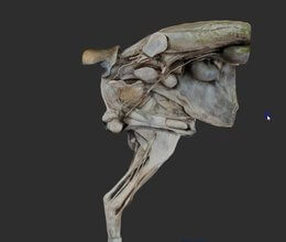
A scan of the male canine urogenital tract, with organs and nerves on one side and the caudal body wall and hindlimb muscles on the other.
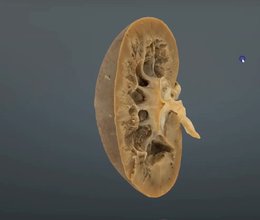
A scanned cut section through the canine kidney, showing the capsule, cortex, medulla and other core structures.
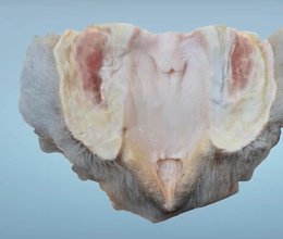
Our canine vestibule content helps you understand the anatomy you need to catheterise a female dog, including the fossa clitoridis.
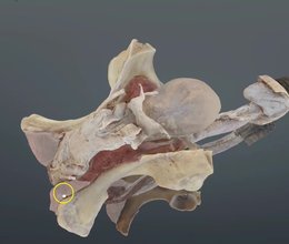
Our scan of the stallion's reproductive tract shows where the accessory sex glands sit and makes sense of the prepuce and its layers.
Ivalalearn
Proud to be part of the Veterinary Information Network family
OUR MISSION:
To shape more confident students and clinicians by creating intuitive, engaging, interactive learning content.
OUR MISSION:
To shape more confident students and clinicians by creating intuitive, engaging, interactive learning content.
Ivala is proud to be a wholly owned subsidiary of the Veterinary Information Network (VIN)
IVALA ® All rights reserved.



