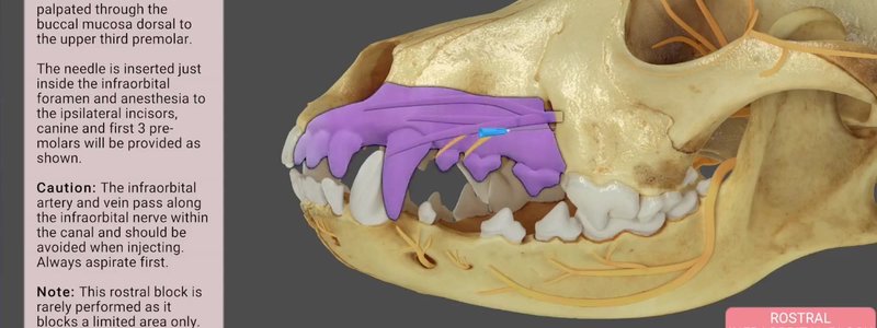
Canine Dental Nerve Blocks in 3D, With Animated Blocks
Our canine dental nerve blocks content animates the area each block anaesthetises and adds technique notes, cautions and the relevant nerves.

Ivalalearn
Proud to be part of the Veterinary Information Network family

These posts look at teeth and the skull in 3D, which is the anatomy you need for dentistry. We cover landmarks, teeth, canals and sinuses in the dog and the horse, as well as the cat, rabbit, guinea pig and chinchilla, where skull and tooth shape are central to dental work in small mammals. There is also a guide to canine dental nerve blocks, shown as animated 3D blocks, so you can follow each nerve to the site of the injection. The posts are written for clinicians and students who want to check a foramen, a tooth root or a sinus before a procedure, and to see how those structures sit together in the head.
Our canine dental nerve blocks content animates the area each block anaesthetises and adds technique notes, cautions and the relevant nerves.
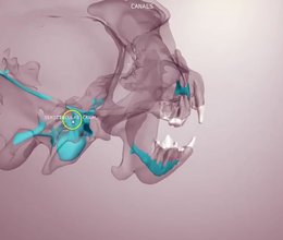
Our canine skull content in four scenes: bony landmarks, teeth and foramina, the canals inside a transparent skull, and a schematic of the cranial nerves.
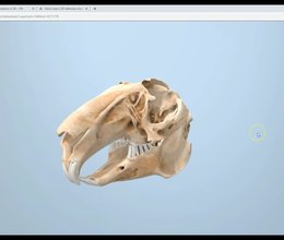
We've released the rabbit skull and dentition in 3D, because so many rabbits present with dental problems and good dentals start with knowing the teeth.
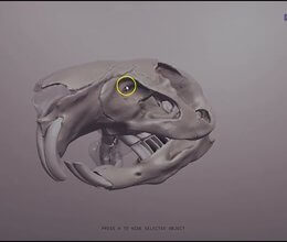
We've added guinea pig and chinchilla skulls so you can learn how the teeth sit relative to one another - handy prep for small-mammal dentistry.
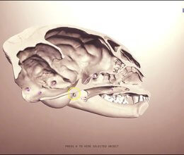
A quick rundown of our feline skull 3D content: teeth with important nerves, bony landmarks, foramina scenes, and Test Yourself mode.
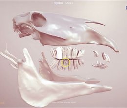
Our interactive equine skull lets you explore how the teeth relate to one another and to the skull, plus a second scene that maps the sinus system for equine dentistry.
Ivalalearn
Proud to be part of the Veterinary Information Network family
OUR MISSION:
To shape more confident students and clinicians by creating intuitive, engaging, interactive learning content.
OUR MISSION:
To shape more confident students and clinicians by creating intuitive, engaging, interactive learning content.
Ivala is proud to be a wholly owned subsidiary of the Veterinary Information Network (VIN)
IVALA ® All rights reserved.



