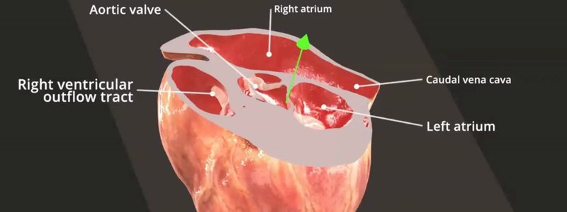
Live Labelling: Labels That Update as You Slice the Heart
Our new live labelling system names anatomical structures on the fly as you slice through the heart, in real time.

Ivalalearn
Proud to be part of the Veterinary Information Network family

These posts help you relate what you see on imaging to the anatomy underneath. We cover the canine heart, including a live section tool and an echocardiography simulator, as well as abdominal radiography and the abdominal and arterial vessels that matter for ultrasound. There is also a guide to our online viewer for microCT studies, first used on canine brain, heart and larynx data. Several posts are written with sonographers and surgeons in mind, so you can follow a vessel or an organ boundary in 3D and then recognise it in a scan. The collection is still weighted towards the dog, which reflects the imaging material we have published so far.
Our new live labelling system names anatomical structures on the fly as you slice through the heart, in real time.
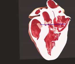
Drag a plane through our 3D canine heart to section it at any position and angle, and explore the chambers and valves inside.
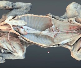
A scanned dissection of a dog revealing the major arterial circulation in situ, from the neck all the way to the median sacral artery.
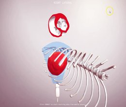
Our proof of concept echo simulator: slice a CT canine heart in any plane with your keyboard or mobile phone sensors, and see the matching 2D image.
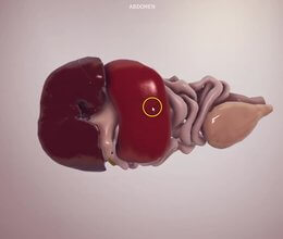
Organs extracted from a real dog CT for accurate topography, plus a radiography scene showing organ outlines as you would see them on X-ray.
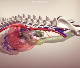
We extracted the vessels from an angiographic CT of a canine abdomen: arterial, venous and portal circulations alongside organs and lymph nodes.
Ivalalearn
Proud to be part of the Veterinary Information Network family
OUR MISSION:
To shape more confident students and clinicians by creating intuitive, engaging, interactive learning content.
OUR MISSION:
To shape more confident students and clinicians by creating intuitive, engaging, interactive learning content.
Ivala is proud to be a wholly owned subsidiary of the Veterinary Information Network (VIN)
IVALA ® All rights reserved.



