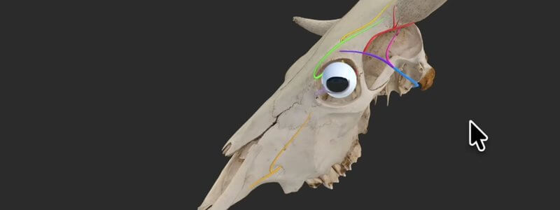
Cow Skull Key Nerves in 3D: Horn and Eye Innervation
Our bovine skull key nerves content highlights the innervation of the horn and eye, a handy review before disbudding, dehorning or eye surgery.

Ivalalearn
Proud to be part of the Veterinary Information Network family

These posts cover ruminant anatomy in 3D, using cow, calf and goat specimens. We look at the ruminant stomachs, including a goat model that we also use when teaching cow and sheep anatomy, the cow skull and the nerves for horn and eye procedures, the bovine stifle, the calf forelimb bones, and a skeleton that also shows the stomach and reproductive tract. There is a surface anatomy guide in both lay and medical terms, which is useful when you are talking with clients. The bovine stifle is close enough to the horse that equine clinicians may find it helpful too. The posts are written for farm animal teaching and for clinical work such as disbudding, imaging and lameness.
Our bovine skull key nerves content highlights the innervation of the horn and eye, a handy review before disbudding, dehorning or eye surgery.
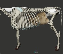
We're giving ruminants some extra love with an interactive 3D cow skeleton, with the stomach and reproductive tract added for context.
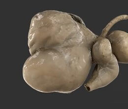
A scan of the ruminant stomachs reconstructed from a real specimen, adding another level of realism to our CT-based schematic.
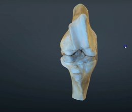
Our scanned, flexed bovine stifle joint adds the distal femur to the proximal tibia, so you can learn the ligaments and how the cruciates attach.
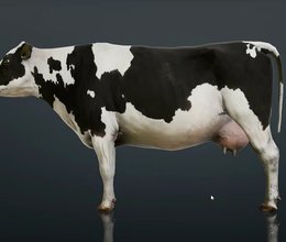
Our bovine surface anatomy covers the major surface landmarks of the cow in both layman and medical terms, ideal for client communication and teaching.
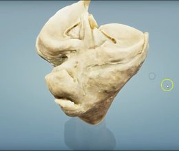
A scan of the proximal tibia of a cow, which our anatomists say is extremely similar to the horse, to help you understand the ligaments of the stifle.
Ivalalearn
Proud to be part of the Veterinary Information Network family
OUR MISSION:
To shape more confident students and clinicians by creating intuitive, engaging, interactive learning content.
OUR MISSION:
To shape more confident students and clinicians by creating intuitive, engaging, interactive learning content.
Ivala is proud to be a wholly owned subsidiary of the Veterinary Information Network (VIN)
IVALA ® All rights reserved.



