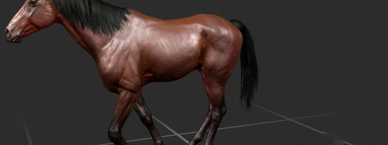
Equine Lameness Gaits in 3D: Grades 1 to 5 at the Walk, Trot and on the Circle
Our animated horse now goes lame: forelimb and hindlimb lameness at grades 1 to 5, at the walk, the trot and on the circle, viewable from any angle.

Ivalalearn
Proud to be part of the Veterinary Information Network family

These posts focus on the hindlimb in 3D. We cover bones, muscles and vessels in the dog, the equine stifle, distal hindlimb and stay apparatus, and the bovine stifle, whose ligaments are also a useful comparison when you are studying the horse. Gait animations are included where they help you relate structure to movement and to lameness examination. The canine posts include musculature sorted by innervation and the bony landmarks you need for orthopaedic work. The aim is to give you a clear picture of the limb from the hip and stifle down to the distal limb, and to support revision as well as clinical examination and surgery.
Our animated horse now goes lame: forelimb and hindlimb lameness at grades 1 to 5, at the walk, the trot and on the circle, viewable from any angle.
Watch our 3D thoroughbred walk, pace, trot, canter on either lead and gallop from any angle, and slow it right down to study each footfall.
Our canine gait animations let you watch a dog walk, amble, pace, trot, canter and gallop, and spin it around to view every stride from any angle.
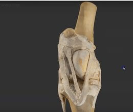
A scan of a real dissected equine stifle, one of the most interesting areas of musculoskeletal anatomy and home of the locking mechanism.
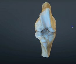
Our scanned, flexed bovine stifle joint adds the distal femur to the proximal tibia, so you can learn the ligaments and how the cruciates attach.
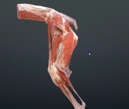
A scan of the canine hindlimb muscles reconstructed from a real cadaver, showing how the muscle bellies are really laid out.
Ivalalearn
Proud to be part of the Veterinary Information Network family
OUR MISSION:
To shape more confident students and clinicians by creating intuitive, engaging, interactive learning content.
OUR MISSION:
To shape more confident students and clinicians by creating intuitive, engaging, interactive learning content.
Ivala is proud to be a wholly owned subsidiary of the Veterinary Information Network (VIN)
IVALA ® All rights reserved.



