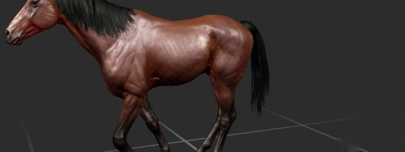
Equine Lameness Gaits in 3D: Grades 1 to 5 at the Walk, Trot and on the Circle
Our animated horse now goes lame: forelimb and hindlimb lameness at grades 1 to 5, at the walk, the trot and on the circle, viewable from any angle.

Ivalalearn
Proud to be part of the Veterinary Information Network family

These posts are for anyone working up lameness or revising the musculoskeletal system. We use 3D models and gait animations to show limb bones, muscles, joints, tendons and ligaments in the dog and the horse, and to help you practise spotting lameness at the walk, trot and on the circle. You will also find guides to the equine distal limb and stay apparatus, canine limb musculature and bony landmarks, and the bovine stifle, which is a useful comparison when you are studying the horse. A calf forelimb post covers centres of ossification in a young farm animal. The material is written so you can move between structure, movement and the clinical examination, rather than treating them as separate topics.
Our animated horse now goes lame: forelimb and hindlimb lameness at grades 1 to 5, at the walk, the trot and on the circle, viewable from any angle.
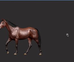
Watch our 3D thoroughbred walk, pace, trot, canter on either lead and gallop from any angle, and slow it right down to study each footfall.
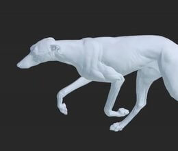
Our canine gait animations let you watch a dog walk, amble, pace, trot, canter and gallop, and spin it around to view every stride from any angle.
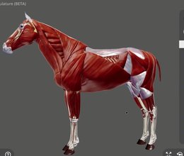
Following our canine full body musculature, the whole musculature of the horse is here, including the trunk and spine muscles we didn't have before.
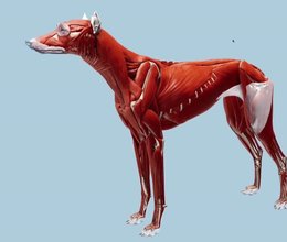
We've brought the entire musculature of the dog together in one 3D model, including the spinal muscles we didn't have before.
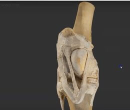
A scan of a real dissected equine stifle, one of the most interesting areas of musculoskeletal anatomy and home of the locking mechanism.
Ivalalearn
Proud to be part of the Veterinary Information Network family
OUR MISSION:
To shape more confident students and clinicians by creating intuitive, engaging, interactive learning content.
OUR MISSION:
To shape more confident students and clinicians by creating intuitive, engaging, interactive learning content.
Ivala is proud to be a wholly owned subsidiary of the Veterinary Information Network (VIN)
IVALA ® All rights reserved.



