
Equine Forelimb From the Elbow Down: Veins, Arteries and Nerves
A scan of the equine forelimb from roughly the elbow down, dissected to reveal the veins, arteries and nerves, colourised to make them easier to see.

Ivalalearn
Proud to be part of the Veterinary Information Network family

A scan of the equine forelimb from roughly the elbow down, dissected to reveal the veins, arteries and nerves, colourised to make them easier to see.
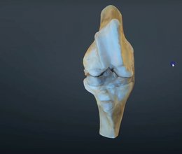
Our scanned, flexed bovine stifle joint adds the distal femur to the proximal tibia, so you can learn the ligaments and how the cruciates attach.
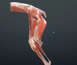
A scan of the canine hindlimb muscles reconstructed from a real cadaver, showing how the muscle bellies are really laid out.
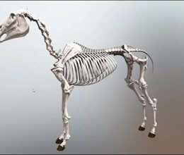
We've expanded our horse content with the equine skeleton, complete with clinical and Latin terms, outline mode, MCQs and free-form self-testing.
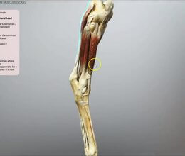
The muscles of the equine distal hindlimb, scanned from a real limb, to give you a much clearer picture of how the flexors and extensors relate.
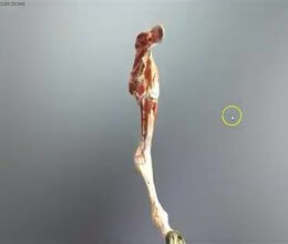
The first of our equine musculature content: the muscles of the equine forelimb, scanned from a real limb for an extremely accurate layout.
Ivalalearn
Proud to be part of the Veterinary Information Network family
OUR MISSION:
To shape more confident students and clinicians by creating intuitive, engaging, interactive learning content.
OUR MISSION:
To shape more confident students and clinicians by creating intuitive, engaging, interactive learning content.
Ivala is proud to be a wholly owned subsidiary of the Veterinary Information Network (VIN)
IVALA ® All rights reserved.



