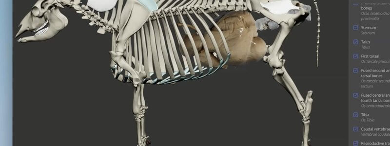
Cow Skeleton in 3D, With the Stomach and Reproductive Tract
We're giving ruminants some extra love with an interactive 3D cow skeleton, with the stomach and reproductive tract added for context.

Ivalalearn
Proud to be part of the Veterinary Information Network family

These posts are about the gastrointestinal tract in 3D. We cover the canine abdomen and liver, including a radiography scene, the equine gut and large intestine, which is useful when you are thinking about colic, and the ruminant stomachs, including a goat specimen that we also use when teaching cow and sheep anatomy. The cow skeleton post brings the stomach in for context alongside the reproductive tract. The aim is to help you make sense of organ position and compartment identity, which matters in abdominal imaging, surgery and clinical examination. Most guides are built from scanned specimens, so you can rotate the tract and see how the parts sit relative to one another.
We're giving ruminants some extra love with an interactive 3D cow skeleton, with the stomach and reproductive tract added for context.
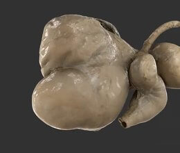
A scan of the ruminant stomachs reconstructed from a real specimen, adding another level of realism to our CT-based schematic.
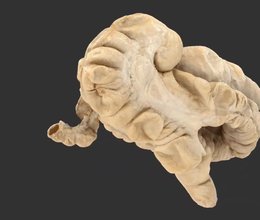
We've added a scan of the equine large intestine from a real specimen, with labelled sacculations and taenial bands, to complement our schematic.
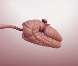
Our equine abdomen content focuses on the gastrointestinal tract, so you can quickly understand the caecum and the different parts of the colon.
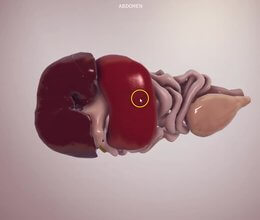
Organs extracted from a real dog CT for accurate topography, plus a radiography scene showing organ outlines as you would see them on X-ray.
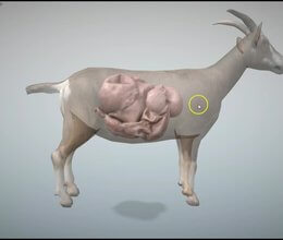
We've released two farm animal scenes built from goat data: the topology of the four stomachs, and the stomachs split open to explore their internal structures.
Ivalalearn
Proud to be part of the Veterinary Information Network family
OUR MISSION:
To shape more confident students and clinicians by creating intuitive, engaging, interactive learning content.
OUR MISSION:
To shape more confident students and clinicians by creating intuitive, engaging, interactive learning content.
Ivala is proud to be a wholly owned subsidiary of the Veterinary Information Network (VIN)
IVALA ® All rights reserved.



