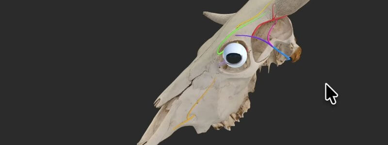
Cow Skull Key Nerves in 3D: Horn and Eye Innervation
Our bovine skull key nerves content highlights the innervation of the horn and eye, a handy review before disbudding, dehorning or eye surgery.

Ivalalearn
Proud to be part of the Veterinary Information Network family

These posts cover the eye, from 3D anatomy to clinical recognition. We walk through the canine eye layer by layer, animate the extraocular muscles and show the effects of nerve lesions, and share a large flashcard deck of clinical images for revision. There is also a cow skull guide that follows the nerves relevant to the eye and to common ocular procedures. The anatomy posts are designed so you can relate a muscle, a nerve or an intraocular structure to the signs you might see in a patient. Taken together, they offer both a spatial understanding of the eye and a way to practise recognising clinical presentations.
Our bovine skull key nerves content highlights the innervation of the horn and eye, a handy review before disbudding, dehorning or eye surgery.
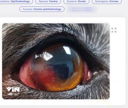
Our canine ophthalmology deck has over 1000 image-based flashcards covering close to every eye condition you might see in dogs.
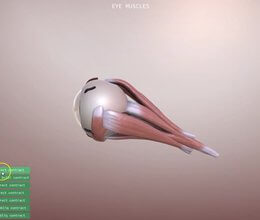
Our new canine eye scene animates what each extrinsic eye muscle does, and simulates the clinical signs of a nerve lesion, starting with the oculomotor nerve.
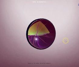
The canine eye in 3D, broken down into its functional anatomy and sortable by anatomical layer, with input from ophthalmologist Ben Blacklock.
Ivalalearn
Proud to be part of the Veterinary Information Network family
OUR MISSION:
To shape more confident students and clinicians by creating intuitive, engaging, interactive learning content.
OUR MISSION:
To shape more confident students and clinicians by creating intuitive, engaging, interactive learning content.
Ivala is proud to be a wholly owned subsidiary of the Veterinary Information Network (VIN)
IVALA ® All rights reserved.



