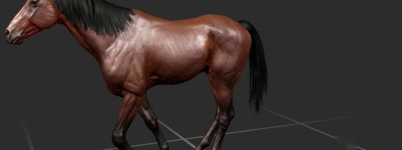
Equine Lameness Gaits in 3D: Grades 1 to 5 at the Walk, Trot and on the Circle
Our animated horse now goes lame: forelimb and hindlimb lameness at grades 1 to 5, at the walk, the trot and on the circle, viewable from any angle.

Ivalalearn
Proud to be part of the Veterinary Information Network family

These posts walk through veterinary anatomy in 3D, using scanned specimens, dissections and animations that you can rotate, section and study at your own pace. We cover bones, muscles, organs, vessels and surface landmarks, mainly in the dog, horse and ruminant, with a handful of posts on the cat and small mammals. Many of the models come from real specimens, so the shapes you see are closer to what you would meet in a dissection room or in practice. A few guides also introduce standard anatomical views and self-testing in the 3D viewer. The aim is to relate what you see on the screen to the anatomy you use in clinics, labs and teaching.
Our animated horse now goes lame: forelimb and hindlimb lameness at grades 1 to 5, at the walk, the trot and on the circle, viewable from any angle.
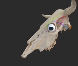
Our bovine skull key nerves content highlights the innervation of the horn and eye, a handy review before disbudding, dehorning or eye surgery.
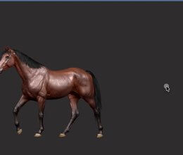
Watch our 3D thoroughbred walk, pace, trot, canter on either lead and gallop from any angle, and slow it right down to study each footfall.
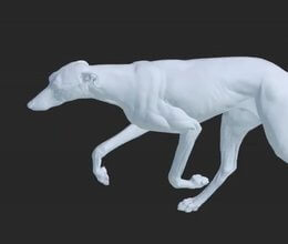
Our canine gait animations let you watch a dog walk, amble, pace, trot, canter and gallop, and spin it around to view every stride from any angle.
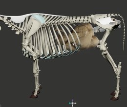
We're giving ruminants some extra love with an interactive 3D cow skeleton, with the stomach and reproductive tract added for context.
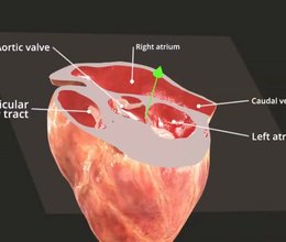
Our new live labelling system names anatomical structures on the fly as you slice through the heart, in real time.
Ivalalearn
Proud to be part of the Veterinary Information Network family
OUR MISSION:
To shape more confident students and clinicians by creating intuitive, engaging, interactive learning content.
OUR MISSION:
To shape more confident students and clinicians by creating intuitive, engaging, interactive learning content.
Ivala is proud to be a wholly owned subsidiary of the Veterinary Information Network (VIN)
IVALA ® All rights reserved.



