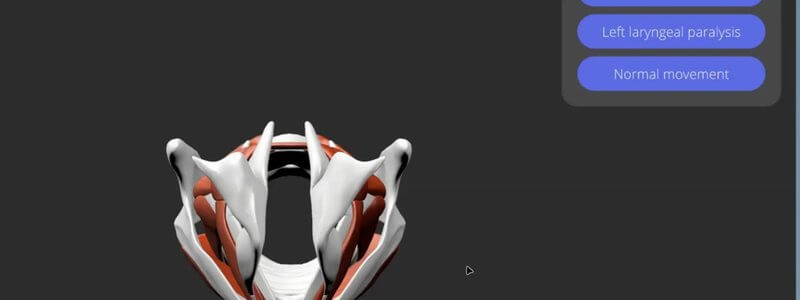
Canine Larynx in 3D, Animated: Normal Movement and Paralysis
Our canine larynx content demystifies the cartilages and muscles, and animates the larynx during normal respiration and laryngeal paralysis.

Ivalalearn
Proud to be part of the Veterinary Information Network family

These posts bring together veterinary neurology, from 3D anatomy to case-based revision. We walk through the canine brain, cranial nerves, middle and inner ear, larynx and autonomic nervous system, and we animate extraocular muscle function and the effects of nerve lesions. Alongside the anatomy there are flashcard decks built from real cases, covering vestibular and cerebellar disease, seizure disorders and videos of abnormal gaits that you can use to practise neurolocalisation. Most of the anatomy is canine, and the case material is drawn from clinical practice. The aim is to help you link a structure, a lesion and the signs you would expect to see, whether you are revising or working up a neurological patient.
Our canine larynx content demystifies the cartilages and muscles, and animates the larynx during normal respiration and laryngeal paralysis.
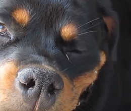
Our vestibular and cerebellar disease deck uses real case videos to help you tell peripheral from central disease and approach each presentation.
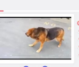
Our neurology case video deck shows you over 50 abnormal gaits from real cases and prompts you to neurolocalise and plan your workup.
Is your patient seizuring, collapsing (or just a bit cuckoo)? Our seizure disorders deck uses real case videos to help you tell the difference.
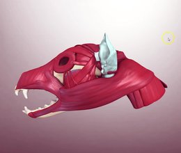
Our canine skull musculature content covers the origins and insertions of the head muscles, lets you sort them by nerve, and has Test Yourself built in.
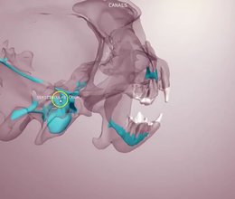
Our canine skull content in four scenes: bony landmarks, teeth and foramina, the canals inside a transparent skull, and a schematic of the cranial nerves.
Ivalalearn
Proud to be part of the Veterinary Information Network family
OUR MISSION:
To shape more confident students and clinicians by creating intuitive, engaging, interactive learning content.
OUR MISSION:
To shape more confident students and clinicians by creating intuitive, engaging, interactive learning content.
Ivala is proud to be a wholly owned subsidiary of the Veterinary Information Network (VIN)
IVALA ® All rights reserved.



