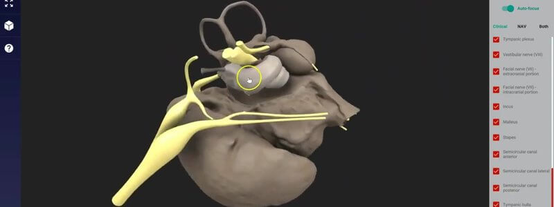
The Canine Middle and Inner Ear in 3D, From a MicroCT Scan
Our canine middle and inner ear, reconstructed from a microCT scan, shows the cavities, ossicles and nerves of the region in remarkable detail.

Ivalalearn
Proud to be part of the Veterinary Information Network family

Our canine middle and inner ear, reconstructed from a microCT scan, shows the cavities, ossicles and nerves of the region in remarkable detail.
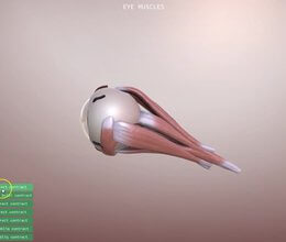
Our new canine eye scene animates what each extrinsic eye muscle does, and simulates the clinical signs of a nerve lesion, starting with the oculomotor nerve.
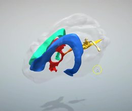
The ventricular system of the canine brain in 3D, with details that are hard to appreciate from textbook diagrams, like the size of the different ducts.
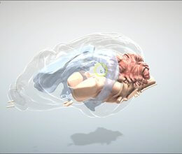
We've added the key structures of the canine brain, from the brainstem and cranial nerves to the forebrain and cerebellum, with clinical neuroanatomy in mind.
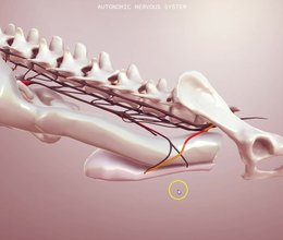
Our 3D schematic of the canine autonomic nervous system shows the sympathetic and parasympathetic outflows, key ganglia and the organs they relate to.
Ivalalearn
Proud to be part of the Veterinary Information Network family
OUR MISSION:
To shape more confident students and clinicians by creating intuitive, engaging, interactive learning content.
OUR MISSION:
To shape more confident students and clinicians by creating intuitive, engaging, interactive learning content.
Ivala is proud to be a wholly owned subsidiary of the Veterinary Information Network (VIN)
IVALA ® All rights reserved.



