
Canine Skull Anatomy in 3D: Landmarks, Teeth, Canals and Cranial Nerves
Here's a tour of our canine skull anatomy content, which is split into four scenes. 💀 We've included two walkthrough videos of it: a quick preview, and a longer one that goes into a little more detail.
Scene 1: bony landmarks
The first scene covers the bony landmarks and prominences of the skull. Rotate it as normal and click on the different landmarks to get relevant information about each one. You can use the grid function on the right to sort through the landmarks, and the search function to find something in particular.
Scene 2: foramina and teeth
In the second scene you can hover over the foramina of the skull to see what each one is. You can also click on the teeth to get more information about them, including their nomenclature. Press the space bar or use the play button to split the objects apart.
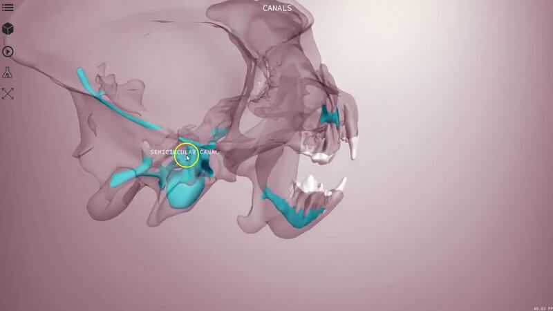
Scene 3: the canals
This one is quite cool. We've made the skull and mandible transparent so you can see the different canals running within the skull, which you can't normally see. Each is coloured differently, including the mandibular canal, and there's a really interesting area at the back with the middle ear cavity and the external acoustic meatus. We've labelled the foramina here too, so you can see how they relate to the canals.
Scene 4: the cranial nerves
The last scene is a schematic of the cranial nerves, mapped out through their relevant foramina, so you can see which nerves run through where. Press the space bar or the play button to swap the skull for the brain and brainstem, and see where the different nerves exit. The easiest way to explore is to hide all the nerves and then pick one, for example the facial nerve or the trigeminal nerve, to see how it runs and read more about it in the top right corner.
Why it's worth the time
Landmarks, foramina and nerve pathways come up again and again, from regional nerve blocks in dentistry to localising a neurological lesion, so it's well worth getting to know them in 3D.
Explore more
FREE for veterinary students, interns, residents & academics at vin.com. Also available by subscription at ivalalearn.com
Want to understand more about the options for accessing the content? Our company FAQs have the answers here pricing, account access and billing.



