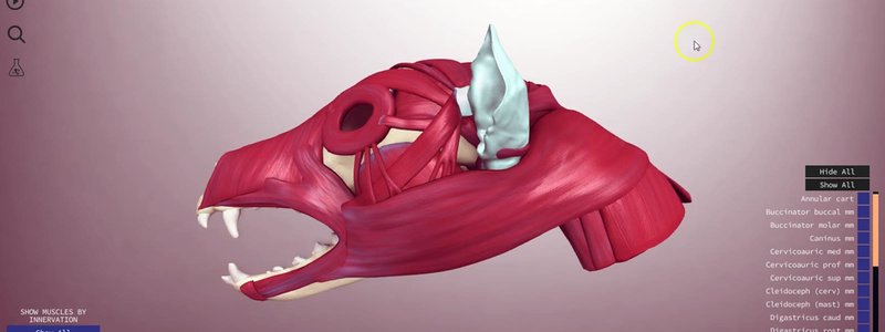
Canine Head Muscles in 3D, Sorted by Innervation
Our canine skull musculature content covers the origins and insertions of the head muscles, lets you sort them by nerve, and has Test Yourself built in.

Ivalalearn
Proud to be part of the Veterinary Information Network family

Our canine skull musculature content covers the origins and insertions of the head muscles, lets you sort them by nerve, and has Test Yourself built in.
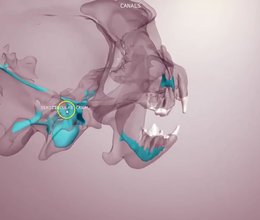
Our canine skull content in four scenes: bony landmarks, teeth and foramina, the canals inside a transparent skull, and a schematic of the cranial nerves.
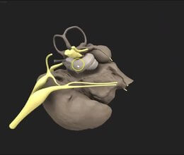
Our canine middle and inner ear, reconstructed from a microCT scan, shows the cavities, ossicles and nerves of the region in remarkable detail.
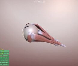
Our new canine eye scene animates what each extrinsic eye muscle does, and simulates the clinical signs of a nerve lesion, starting with the oculomotor nerve.
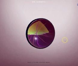
The canine eye in 3D, broken down into its functional anatomy and sortable by anatomical layer, with input from ophthalmologist Ben Blacklock.
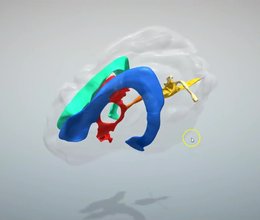
The ventricular system of the canine brain in 3D, with details that are hard to appreciate from textbook diagrams, like the size of the different ducts.
Ivalalearn
Proud to be part of the Veterinary Information Network family
OUR MISSION:
To shape more confident students and clinicians by creating intuitive, engaging, interactive learning content.
OUR MISSION:
To shape more confident students and clinicians by creating intuitive, engaging, interactive learning content.
Ivala is proud to be a wholly owned subsidiary of the Veterinary Information Network (VIN)
IVALA ® All rights reserved.



