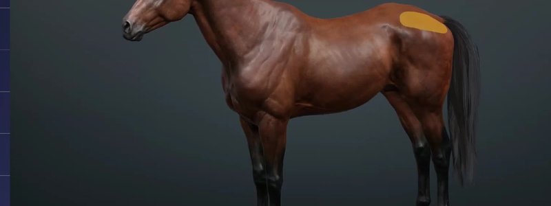
Equine Surface Anatomy in 3D: Speak Your Clients' Language
Our equine surface anatomy covers the major surface regions of the horse with a focus on layman terms, so you and your clients speak the same language.

Ivalalearn
Proud to be part of the Veterinary Information Network family

Our equine surface anatomy covers the major surface regions of the horse with a focus on layman terms, so you and your clients speak the same language.
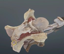
Our scan of the stallion's reproductive tract shows where the accessory sex glands sit and makes sense of the prepuce and its layers.
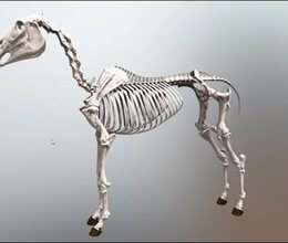
We've expanded our horse content with the equine skeleton, complete with clinical and Latin terms, outline mode, MCQs and free-form self-testing.
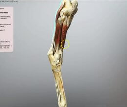
The muscles of the equine distal hindlimb, scanned from a real limb, to give you a much clearer picture of how the flexors and extensors relate.
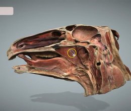
We've added a scanned median section of the equine head, updated the sinus terminology to match the NAV, and made navigation smoother.
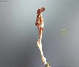
The first of our equine musculature content: the muscles of the equine forelimb, scanned from a real limb for an extremely accurate layout.
Ivalalearn
Proud to be part of the Veterinary Information Network family
OUR MISSION:
To shape more confident students and clinicians by creating intuitive, engaging, interactive learning content.
OUR MISSION:
To shape more confident students and clinicians by creating intuitive, engaging, interactive learning content.
Ivala is proud to be a wholly owned subsidiary of the Veterinary Information Network (VIN)
IVALA ® All rights reserved.



