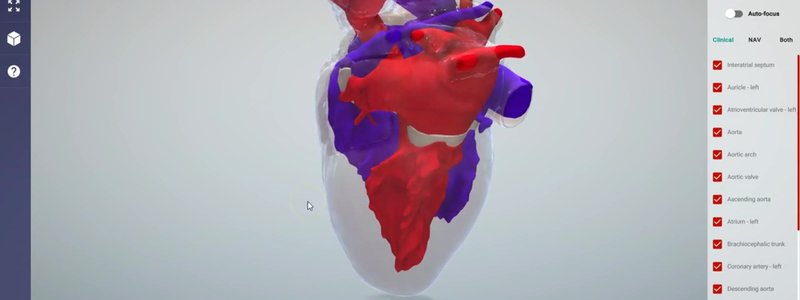
Canine Heart in 3D: Surface Anatomy and Internal Circulations
We've added the surface anatomy of the canine heart, and updated its internal anatomy to show the left and right sides of the circulation.

Ivalalearn
Proud to be part of the Veterinary Information Network family

We've added the surface anatomy of the canine heart, and updated its internal anatomy to show the left and right sides of the circulation.
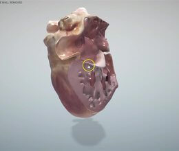
Our canine chest content now includes the heart with the right ventricular wall removed, so you can explore the papillary muscles, chordae tendineae and more.
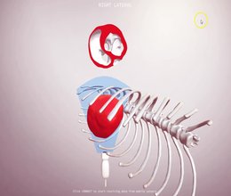
Our proof of concept echo simulator: slice a CT canine heart in any plane with your keyboard or mobile phone sensors, and see the matching 2D image.
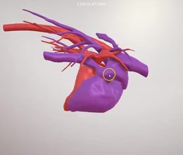
A walkthrough of our canine chest content: lung fields, the major vessels, the left and right circulations from a CT, and the bronchial tree.
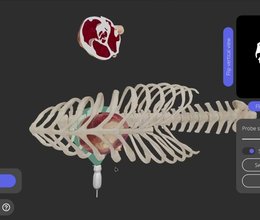
Our canine echocardiography simulator just got heart valves, a more realistic probe starting point and thumb-pivot fanning. Here's what's new and why it matters when you scan a real patient.
Ivalalearn
Proud to be part of the Veterinary Information Network family
OUR MISSION:
To shape more confident students and clinicians by creating intuitive, engaging, interactive learning content.
OUR MISSION:
To shape more confident students and clinicians by creating intuitive, engaging, interactive learning content.
Ivala is proud to be a wholly owned subsidiary of the Veterinary Information Network (VIN)
IVALA ® All rights reserved.



