
Canine Heart in 3D: Surface Anatomy and Internal Circulations
We've updated our canine chest project with two new pieces of heart content: the surface anatomy of the heart and its internal circulations. ❤️
Surface anatomy of the heart
The first new scene covers the surface anatomy of the canine heart. You can interact with the different aspects of the surface to get more information about each one.
The left and right circulations
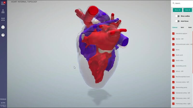
We've also updated the internal anatomy of the heart to show the left and right sides of the circulation. The left side is in red to represent oxygenated blood, and the right side is in blue to represent deoxygenated blood. Seeing the two sides together is a great way to understand how blood moves through the heart.
As usual, you can interact with the objects and hide them. In the video we hide the left atrium to get a better look at the left atrioventricular valve, then hide the ventricle too.
All the normal features
- Switch to Latin terminology, or show both clinical and Latin terms together
- Create your own custom tests with our testing system, either MCQs or Type Yourself
Have a play
Have a play with it, enjoy it, and let us know what you think.
Explore more
FREE for veterinary students, interns, residents & academics at vin.com. Also available by subscription at ivalalearn.com
Want to understand more about the options for accessing the content? Our company FAQs have the answers here pricing, account access and billing.



