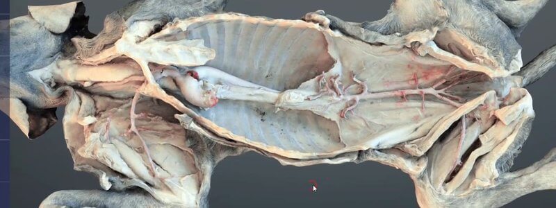
Canine Arterial Vasculature in 3D, From Neck to Sacrum
A scanned dissection of a dog revealing the major arterial circulation in situ, from the neck all the way to the median sacral artery.

Ivalalearn
Proud to be part of the Veterinary Information Network family

A scanned dissection of a dog revealing the major arterial circulation in situ, from the neck all the way to the median sacral artery.
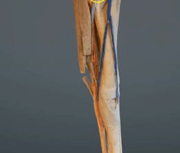
A scan of the equine forelimb from roughly the elbow down, dissected to reveal the veins, arteries and nerves, colourised to make them easier to see.
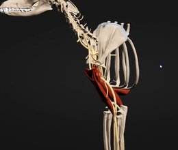
Our canine brachial plexus content was built to help teach the RUMM block, and it's also a great overview of the nerves of the forelimb.
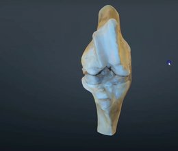
Our scanned, flexed bovine stifle joint adds the distal femur to the proximal tibia, so you can learn the ligaments and how the cruciates attach.
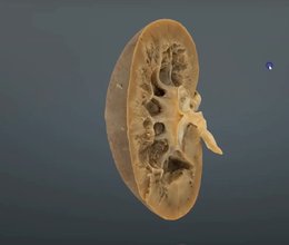
A scanned cut section through the canine kidney, showing the capsule, cortex, medulla and other core structures.
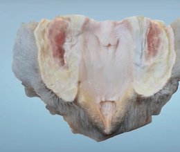
Our canine vestibule content helps you understand the anatomy you need to catheterise a female dog, including the fossa clitoridis.
Ivalalearn
Proud to be part of the Veterinary Information Network family
OUR MISSION:
To shape more confident students and clinicians by creating intuitive, engaging, interactive learning content.
OUR MISSION:
To shape more confident students and clinicians by creating intuitive, engaging, interactive learning content.
Ivala is proud to be a wholly owned subsidiary of the Veterinary Information Network (VIN)
IVALA ® All rights reserved.



