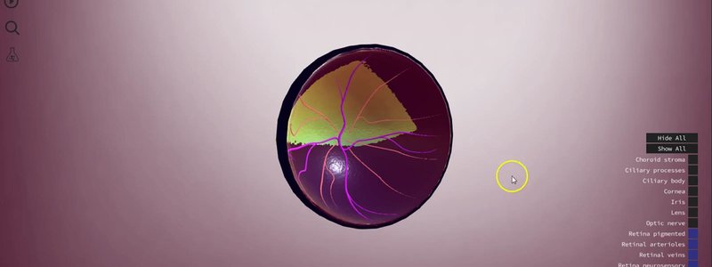
The Canine Eye in 3D: Functional Anatomy Layer by Layer
The canine eye in 3D, broken down into its functional anatomy and sortable by anatomical layer, with input from ophthalmologist Ben Blacklock.

Ivalalearn
Proud to be part of the Veterinary Information Network family

The canine eye in 3D, broken down into its functional anatomy and sortable by anatomical layer, with input from ophthalmologist Ben Blacklock.
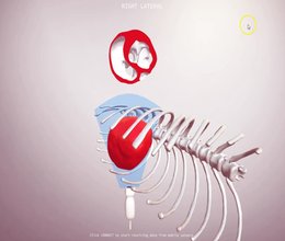
Our proof of concept echo simulator: slice a CT canine heart in any plane with your keyboard or mobile phone sensors, and see the matching 2D image.
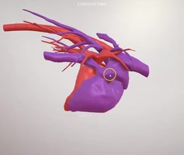
A walkthrough of our canine chest content: lung fields, the major vessels, the left and right circulations from a CT, and the bronchial tree.
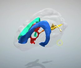
The ventricular system of the canine brain in 3D, with details that are hard to appreciate from textbook diagrams, like the size of the different ducts.
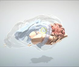
We've added the key structures of the canine brain, from the brainstem and cranial nerves to the forebrain and cerebellum, with clinical neuroanatomy in mind.
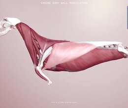
The body wall muscles are a tricky area to picture. Our 3D scene shows how the layers sit in relation to one another, with information on every muscle.
Ivalalearn
Proud to be part of the Veterinary Information Network family
OUR MISSION:
To shape more confident students and clinicians by creating intuitive, engaging, interactive learning content.
OUR MISSION:
To shape more confident students and clinicians by creating intuitive, engaging, interactive learning content.
Ivala is proud to be a wholly owned subsidiary of the Veterinary Information Network (VIN)
IVALA ® All rights reserved.



