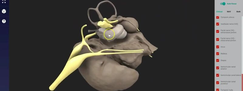
The Canine Middle and Inner Ear in 3D, From a MicroCT Scan
Our canine middle and inner ear, reconstructed from a microCT scan, shows the cavities, ossicles and nerves of the region in remarkable detail.

Ivalalearn
Proud to be part of the Veterinary Information Network family

Our canine middle and inner ear, reconstructed from a microCT scan, shows the cavities, ossicles and nerves of the region in remarkable detail.
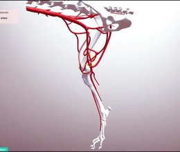
Our limb vasculature content now includes the arteries and veins of the canine hindlimb, plus a new switch between clinical and Latin (NAV) terminology.
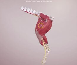
Understand the origins, insertions and relations of the canine hindlimb muscles in 3D, sort them by nerve supply, and explore the canine stifle.
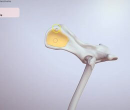
Our canine hindlimb bones update: select each individual bone, see entire landmark regions highlight, and use autofocus as you move down the leg.
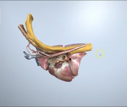
We've added the canine right hemithorax to the Superproject, great for following the oesophagus, and made selected objects highlight in bright orange.
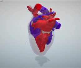
We've added the surface anatomy of the canine heart, and updated its internal anatomy to show the left and right sides of the circulation.
Ivalalearn
Proud to be part of the Veterinary Information Network family
OUR MISSION:
To shape more confident students and clinicians by creating intuitive, engaging, interactive learning content.
OUR MISSION:
To shape more confident students and clinicians by creating intuitive, engaging, interactive learning content.
Ivala is proud to be a wholly owned subsidiary of the Veterinary Information Network (VIN)
IVALA ® All rights reserved.



